Understanding Rayscan Alpha Plus Benefits
The Rayscan Alpha Plus is an advanced imaging system used in modern dental practices for enhanced diagnostic capabilities. Known for its precision and innovative technology, this equipment provides clear images critical for accurate diagnoses and treatment planning. This article explores its features, benefits, and common questions surrounding its use in the dental industry.
Introduction to Dental Imaging Technology
In the evolving landscape of dental health, advanced imaging technology plays a pivotal role in enhancing diagnostic accuracy and treatment outcomes. The Rayscan Alpha Plus stands out as a hallmark of innovation and precision in this domain, offering dental professionals the tools needed to make informed decisions. The integration of technology in dentistry has transformed the way practitioners diagnose and treat conditions, making it possible to visualize structures in ways that were previously unimaginable. This not only encompasses the act of seeing inside the mouth but extends to understanding the complex relationships between structures that affect patient health.
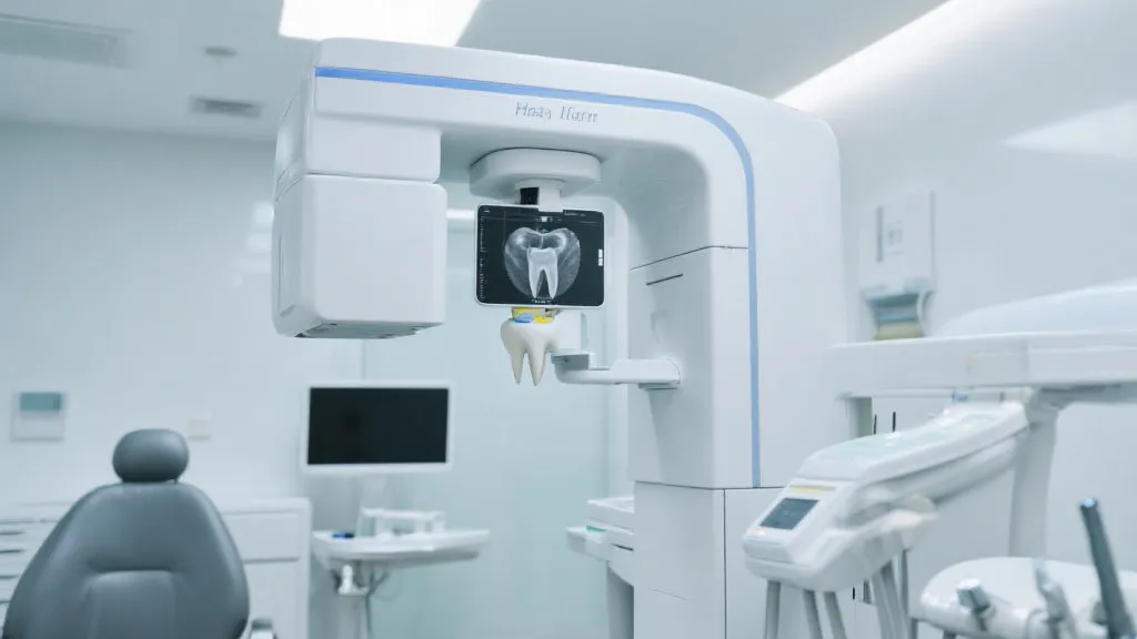
What is the Rayscan Alpha Plus?
The Rayscan Alpha Plus is a top-of-the-line dental imaging system designed to provide high-definition images using advanced cone-beam computed tomography (CBCT) technology. This system is renowned for its ability to produce detailed 3D images, which are invaluable in diagnosing a range of dental conditions and planning complex procedures. The Rayscan Alpha Plus utilizes a rotating x-ray source and a digital receptor to capture multiple images in a single sweep, consolidating the effort into a comprehensive scan. With the proliferation of dental surgeries and the need for higher fidelity in imaging quality, systems like the Rayscan Alpha Plus herald a new era in digital dentistry. This technology enables practitioners not merely to diagnose existing issues but to anticipate potential problems as well.
Key Features of the Rayscan Alpha Plus
- High Resolution Imaging: The system captures images with exceptional clarity, allowing for detailed examination of dental structures. This feature facilitates new levels of investigation into anatomical structures, enabling practitioners to detect subtle pathologies that might go unnoticed with conventional imaging.
- Low Radiation Exposure: Employing effective dose optimization techniques ensures patient safety without compromising image quality. The Rayscan Alpha Plus uses innovative algorithms that adjust the radiation dose based on the patient's size and the area being scanned, significantly minimizing risks associated with radiation exposure.
- Flexible Imaging Options: Offers a full range of scanning field sizes, catering to various diagnostic requirements from full maxillofacial scans to focused area imaging. The ability to customize the field of view ensures that practitioners can obtain the requisite detail for any given procedure, enhancing their treatment options.
Benefits of Using the Rayscan Alpha Plus
The primary benefits of this cutting-edge imaging system include:
- Enhanced Diagnostic Accuracy: Precise images aid in accurate diagnosis of dental issues, facilitating better treatment planning and outcomes. The Rayscan Alpha Plus excels in providing multi-dimensional visualizations of complex cases, which is vital when planning interventions such as implants, extractions, and reconstructive surgeries.
- Improved Patient Experience: Faster image acquisition and reduced need for multiple scans contribute to a more comfortable and efficient patient experience. With streamlined workflows, dental professionals can significantly cut down on patient wait times and ease anxiety associated with imaging procedures.
- Comprehensive Treatment Planning: Detailed 3D models assist dentists in visualizing and planning surgeries and other treatment modalities with greater precision. By using simulations and models derived from the captured images, dentists can discuss treatment options with patients more effectively, providing insights into what the patient can expect.
Comparative Table of Dental Imaging Systems
| Feature | Rayscan Alpha Plus | Competitor X | Competitor Y |
|---|---|---|---|
| Imaging Technology | CBCT | Panoramic X-Ray | CBCT |
| Radiation Dosage | Low | Medium | High |
| Field of View | Adjustable | Fixed | Adjustable |
| Cost-effectiveness | High | Medium | Low |
| 3D Visualization | Available | Not Available | Available |
| Software Integration | Full Integration with Practice Management Software | Limited Integration | Moderate Integration |
Frequently Asked Questions (FAQs)
1. How does the Rayscan Alpha Plus enhance treatment accuracy?
The detailed 3D images generated by the Rayscan Alpha Plus allow for precise mapping and diagnosis, which is crucial for tailoring an effective treatment plan. Moreover, with the help of advanced imaging, dentists can pinpoint the exact location of anatomical structures to avoid damaging nerves and blood vessels during procedures.
2. Is the radiation exposure safe for patients?
Yes, the Rayscan Alpha Plus is designed to use minimal radiation while maintaining high image quality, ensuring patient safety during scans. The low exposure is particularly significant for vulnerable populations, such as children or patients requiring frequent imaging, where cumulative radiation dose is a concern.
3. Who typically uses the Rayscan Alpha Plus?
This imaging system is widely used by dental professionals, including orthodontists, oral surgeons, and periodontists, who require detailed imaging for advanced procedures. Its use is also expanding into fields such as implantology and endodontics, where precise imaging is crucial for successful outcomes.
4. Can the system handle imaging for pediatric patients?
Yes, the system’s adjustable settings make it suitable for children, allowing for safe and accurate imaging that caters to smaller anatomies. This feature not only bolsters patient safety but also enables pediatric dentists to accurately monitor dental development and plan treatments accordingly.
The Role of Advanced Imaging in Dental Specialties
The integration of advanced imaging devices like the Rayscan Alpha Plus has significantly impacted various dental specialties. For instance, in orthodontics, clear visualization of the patient’s dental and skeletal anatomy can greatly influence treatment trajectory. Rather than relying on conventional two-dimensional images, orthodontists can use 3D representations to assess relationships between teeth and bone structures and develop personalized treatment plans.
In oral surgery, precise imaging is indispensable. Surgeons can leverage the 3D models produced by the Rayscan Alpha Plus to enhance their surgical planning. By visualizing the patient's anatomy from multiple angles, they can anticipate challenges, streamline procedures, and minimize both operative and postoperative complications. Surgical guides produced from these imaging scans further improve the accuracy of complex procedures like implant placements.
Periodontists are also able to utilize the advantages of high-resolution imaging. Understanding the extent of periodontal disease is fundamentally alterable by the insights offered through three-dimensional imaging. By visualizing bone levels, infections, and the positioning of teeth relative to the surrounding structures, treatment strategies can be tailored to individual patient needs.
Integration of Imaging Technology with Digital Dentistry
The Rayscan Alpha Plus serves as a cornerstone in the broader framework of digital dentistry—a concept that encompasses the use of digital technologies to improve dental care efficiency and outcomes. With digital workflows, radiographic images from the Rayscan Alpha Plus can be easily integrated into patient records and treatment planning software. This seamless integration allows dentists to manage patient information effectively and collaborate with team members more efficiently.
The advent of digital impressions has further propelled innovation in the field. The coupling of 3D imaging from devices like the Rayscan Alpha Plus with intraoral scanners means that dental professionals can achieve a comprehensive understanding of the patient's mouth in real time. This confluence of technologies facilitates more precise treatment options, reduces procedural time, and enhances the overall patient experience.
Advancements in Dental Imaging Technology
While the Rayscan Alpha Plus is at the forefront of current dental imaging technology, the field continues to evolve with advancements that are pushing the boundaries further. Emerging trends in dental imaging technology include artificial intelligence (AI), which is being increasingly incorporated into radiographic analysis. AI algorithms can assist radiologists in interpreting images, highlighting areas of concern that may require closer examination. This can significantly aid in the early detection of conditions such as dental caries and cancers, which can often be missed in a standard evaluation.
Another area of growth is the move toward cone-beam computed tomography (CBCT) systems that are becoming more compact and user-friendly. As manufacturers focus on designing systems that save space while enhancing imaging capabilities, dental practices with limited space can still benefit from high-quality imaging technology. Furthermore, advancements in wireless technology are enabling dental imaging systems to operate without cumbersome wiring, facilitating ease of use and enhancing operational efficiency in busy clinical environments.
Addressing Patient Comfort and Anxiety with Dental Imaging
The scanning process has undergone significant modifications as a result of technological advancements. The Rayscan Alpha Plus has taken into consideration the apprehensions that many patients experience concerning dental visits. By minimizing the duration of scans and the complexity of the procedure, patients find themselves more at ease. In addition, innovations in imaging technology have made it possible to design systems that are quieter and less intimidating, creating a welcoming atmosphere.
Some clinics even incorporate patient education into their imaging process. By allowing patients to visualize their scans in real-time, practitioners can demystify the process and remove fears associated with the unknown. The Rayscan Alpha Plus can display scans immediately on a screen, providing dentists the opportunity to explain findings to patients on-the-spot, bridging communication gaps and fostering informed consent.
Future Directions in Dental Imaging
The future of dental imaging technology appears poised for remarkable advancements. Research is ongoing into improving imaging resolution, reducing scan times, and continually optimizing radiation doses to ensure patient safety. The increasing emphasis on preventative care in dentistry is also directing attention toward imaging technologies that facilitate earlier interventions through enhanced diagnostic tools.
Wearable imaging technology may also emerge as a game-changer in the dental field. Innovations will likely lead to devices that can be utilized for continual monitoring of dental conditions, allowing patients to receive real-time data regarding their oral health. Such technology could empower patients to make better health decisions and keep dental practitioners informed regarding their patients’ conditions between visits.
Conclusion
The Rayscan Alpha Plus represents the forefront of dental imaging technology, providing unmatched precision and safety in clinical settings. For dental practices aiming to elevate their diagnostic and treatment planning capabilities, investing in this advanced equipment is a strategic decision. Ultimately, the Rayscan Alpha Plus not only enhances clinical outcomes but also improves patient care and experience. By embracing this technology, dental practitioners can pave the way for a new standard of care that prioritizes accuracy, safety, and patient satisfaction. As the field continues to progress, these imaging systems are set to redefine the boundaries of what is possible in dental diagnostics and treatment.