Revolutionizing Dental Imaging Technology
The Rayscan Alpha Plus is at the forefront of dental imaging innovation, providing unparalleled diagnostic capabilities in dental clinics worldwide. This high-tech device leverages cutting-edge technology to enhance patient care by providing precise and detailed imaging, crucial for accurate diagnosis and treatment planning. This review delves into its technical advantages and practical applications.
Introduction to Dental Imaging Technology
In the realm of modern dentistry, advanced imaging technology plays a crucial role in ensuring accurate diagnoses and effective treatment plans. Leading the charge is the Rayscan Alpha Plus, a sophisticated piece of equipment that stands out in the dental imaging market. This article explores its impact, benefits, and why it is a preferred choice among dental professionals. With the continual evolution in technology, the Rayscan Alpha Plus integrates cutting-edge features that ultimately enhance clinical decision-making and improve patient outcomes. As dental practices adapt to these advancements, understanding the significance of systems like the Rayscan Alpha Plus becomes increasingly pertinent.
Understanding the Rayscan Alpha Plus
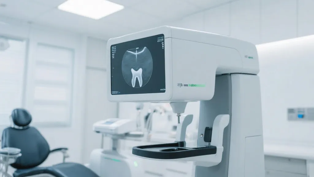
The Rayscan Alpha Plus is a state-of-the-art dental imaging device known for its exceptional image quality and user-friendly interface. Designed with the latest technology, it helps clinicians make swift, precise diagnostics—a factor that significantly enhances patient care. This device incorporates 3D imaging capabilities, making it invaluable for procedures requiring a detailed view of the oral structure. The instrument provides comprehensive imaging, including panoramic, cephalometric, and cone beam computed tomography (CBCT), thereby equipping dentists with the necessary tools to address various clinical situations accurately.
Technical Advantages
One of the primary benefits of the Rayscan Alpha Plus is its ability to produce high-resolution images that assist in pinpointing dental issues with unmatched precision. Its innovative software ensures minimal radiation exposure, prioritizing patient safety while delivering high-quality images. This balance of safety and efficiency marks a significant leap forward in dental imaging technology. The device's ability to generate images from different angles leads to a three-dimensional visualization of the anatomy, which is highly critical for diagnosing complex conditions such as impaction or anomalies in tooth development. Moreover, advanced algorithms within the device enhance contrast and clarity, allowing for better examination of bone structures and soft tissues surrounding the teeth.
Applications in Dentistry
From routine check-ups to complex dental surgeries, the Rayscan Alpha Plus proves invaluable across various dental specialties. Its capability to visualize intricate details makes it indispensable for orthodontists to assess and plan treatments accurately. With the precision that the Rayscan Alpha Plus offers, orthodontists can routinely track the progression of treatment, facilitating adjustments and improving patient satisfaction. Periodontists and maxillofacial surgeons also benefit from its detailed imaging, which aids in surgical planning and monitoring of healing post-procedures. The ability to take a comprehensive view of the maxillofacial region allows for better planning in implants and tooth extractions. For endodontists, the clarity of the internal tooth structures enhances the diagnosis of root canal complications, ultimately leading to more effective treatment solutions.
Furthermore, the Rayscan Alpha Plus provides features like real-time imaging. This capability assists practitioners in making immediate clinical decisions during procedures, reducing the need for multiple visits and enhancing overall patient experience. Whether it’s placing a dental implant or performing a root canal, the resultant images can facilitate decisions with an unprecedented level of confidence.
User Experience and Accessibility
The intuitive design of the Rayscan Alpha Plus allows for ease of use, enabling dental professionals to focus more on patient interaction and care rather than on navigating complex machinery. This user-friendly approach decreases the learning curve, allowing practices to integrate the device into their daily operations swiftly. Its streamlined operability ensures consistent and reliable results, enhancing workflow efficiency. The graphical user interface is designed to guide users from initial setup to imaging in simple steps, which ensures that less time is spent training staff. Additionally, the ability to store images digitally within the system allows for easy retrieval and sharing with patients, fostering better communication.
As dental technology continues to evolve, the Rayscan Alpha Plus also embraces upgrades and modifications through software updates. Such adaptability allows users to keep pace with advancements in dental imaging without substantial additional investment, demonstrating a commitment to maintaining the latest practices in dental care. Moreover, the device’s connective capabilities enable integration with practice management software, resulting in streamlined workflows and improved patient record management.
Table: Comparing Dental Imaging Technologies
| Feature | Rayscan Alpha Plus | Conventional X-ray Machines |
|---|---|---|
| Image Quality | High-Resolution 3D Imaging | 2D Imaging |
| Radiation Exposure | Minimal | Higher |
| User Interface | Intuitive and User-friendly | Basic |
| Versatility | Multi-disciplinary Applications | Limited to Basic Diagnostics |
| Real-Time Imaging | Yes | No |
| Digital Storage | Cloud-Compatible Storage Options | Physical Film Storage |
| Cost of Maintenance | Lower with Software Updates | Higher due to Film and Development |
Frequently Asked Questions
- What sets the Rayscan Alpha Plus apart from other imaging devices?
The combination of high-resolution 3D imaging capabilities and minimal radiation exposure sets it apart. Additionally, the adaptability and range of applications make it a versatile choice for dental professionals.
- Is the device suitable for all dental practices?
Yes, its versatility makes it ideal for diverse dental specialties and procedures. Whether a practice is focused on restorative dentistry, orthodontics, or oral surgery, the Rayscan Alpha Plus can cater to those needs.
- How does it enhance patient care?
By providing accurate and detailed imaging, it aids in precise diagnosis and effective treatment planning. This leads to improved patient outcomes, increased trust in the dentist, and an overall better patient experience.
- Does the Rayscan Alpha Plus require extensive training to operate?
The user-friendly interface and design significantly reduce the training time for new operators. Most dental professionals can become adept at using the device with minimal guidance.
- How frequently can the device be upgraded?
The software of the Rayscan Alpha Plus can be updated regularly, allowing for improved functionality and features that keep pace with advancements in dental imaging technology.
- Can the images be easily shared with patients and specialists?
Yes, the digital storage capabilities enable easy sharing of images with both patients and other dental specialists, facilitating more coordinated patient care.
Clinical Case Studies
Understanding the practical applications and effectiveness of the Rayscan Alpha Plus in real-world settings can further justify its adoption in dental practices. Below are several clinical case studies that illustrate its advantages during different dental procedures.
Case Study 1: Orthodontic Treatment Planning
An orthodontic practice utilized the Rayscan Alpha Plus to evaluate a patient presenting with a complex malocclusion. Using the device, the orthodontist obtained high-resolution 3D images of the patient's jaws and teeth, revealing impaction of several unerupted teeth. These details allowed for a tailored treatment plan that incorporated extractions and the use of braces. Throughout the treatment, the orthodontist used the device to monitor progress, making adjustments as necessary based on the detailed views provided. The patient reported high satisfaction with the treatment outcomes, attributing much to the clarity and detail offered by the Rayscan Alpha Plus.
Case Study 2: Surgical Implant Planning
A team of oral surgeons integrated the Rayscan Alpha Plus into their surgical planning protocols for dental implant placement. In one particular case, a patient required an implant in the posterior maxilla, a region known for its proximity to vital anatomical structures. The 3D imaging capabilities allowed the surgical team to visualize the sinus cavity and assess the density of bone in different areas, leading to a strategic implant placement that minimized risks during surgery. Post-operatively, follow-up imaging using the same device confirmed the correct positioning and integration of the implant into the bone structure.
Case Study 3: Endodontic Diagnosis
In a dental practice specializing in endodontics, a patient presented with persistent discomfort following a previous root canal treatment. The practitioner utilized the Rayscan Alpha Plus to image the anatomical complexities of the tooth’s root canal system, which are often not evident in 2D images. The 3D visualization revealed a missed canal that was the likely source of infection. As a result, the clinician adjusted the treatment plan to incorporate additional cleaning and sealing of the previously untreated canal. This ultimately led to the resolution of the patient’s discomfort.
The Future of Dental Imaging
The advancement of dental imaging technology is expected to continue at a rapid pace. Emerging technologies such as artificial intelligence (AI) and machine learning are beginning to influence how dental imaging interprets and analyzes images. Future enhancements may include automated detection of common dental issues within images, which could expedite the diagnostic process and enhance treatment planning. As these technologies continue to evolve, devices like the Rayscan Alpha Plus will remain at the forefront, adapting to incorporate new findings and tools, ensuring that dental professionals can provide the best care for their patients.
Furthermore, the emphasis on minimally invasive procedures is anticipated to rise, aligning with patient preferences for treatments that preserve natural tooth structure and minimize discomfort. The roles of imaging technologies will expand in relation to minimally invasive techniques, serving as vital tools for planning and executing such procedures flawlessly. Continuous patient education regarding the benefits of advanced imaging will also bolster demand as patients become more informed and proactive about their oral health.
Conclusion
As dental practices aim for improved patient outcomes, integrating advanced technology like the Rayscan Alpha Plus is imperative. This device not only enhances diagnostic accuracy and treatment efficacy but also prioritizes patient safety and comfort. It exemplifies where modern dentistry is headed—toward tech-driven, patient-centered care. The combination of versatile applications, high-resolution imagery, and intuitive operation reflects a significant evolution in dental practice management and patient care approaches. Embracing this technology is essential for dental professionals committed to elevating their practice to meet the demands of today's patients and complex dental conditions.